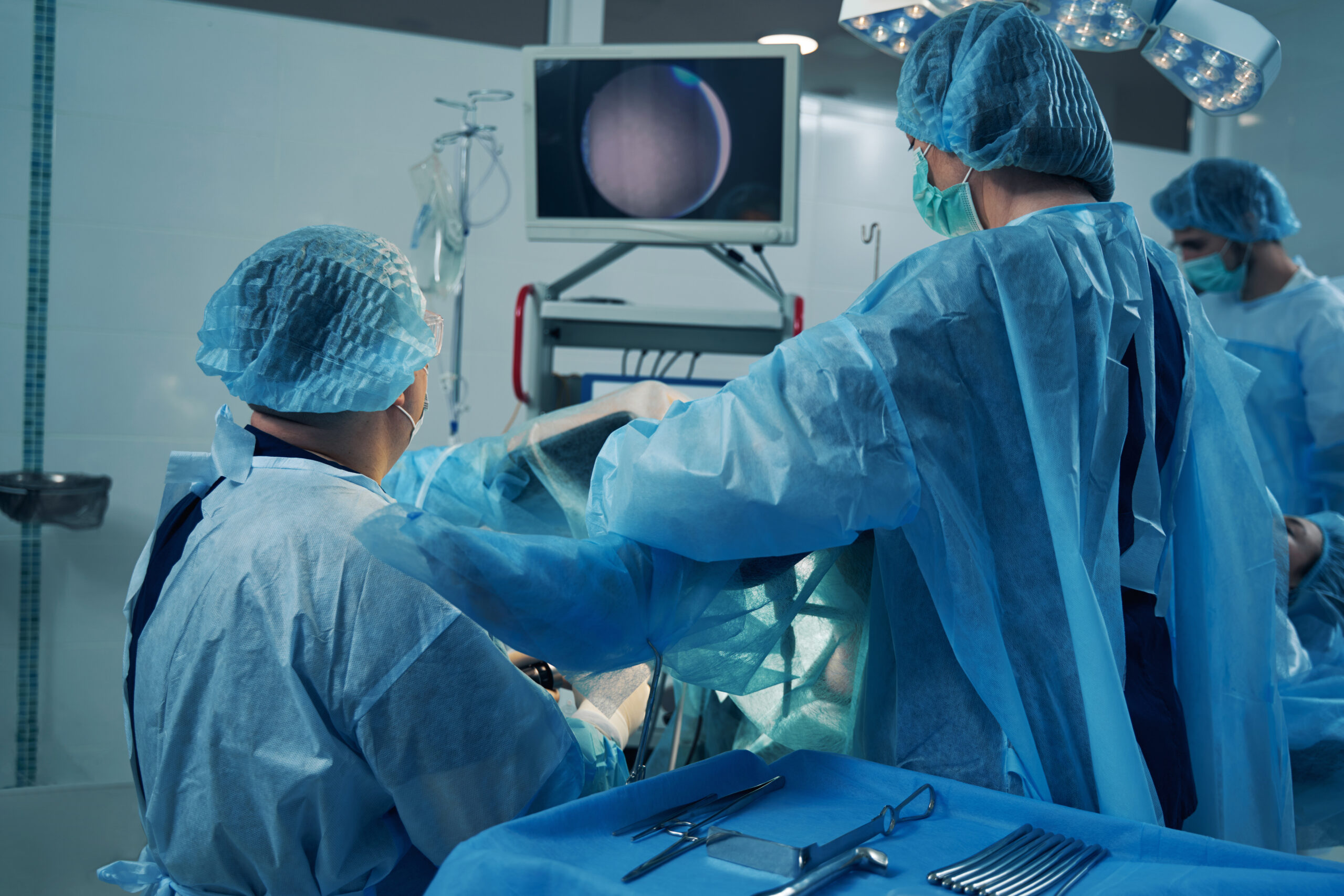
Hysteroscopy (H/S) is a minimally invasive diagnostic and surgical procedure used to visualize the inside of the uterus and diagnose or treat various gynecological conditions. It involves the insertion of a thin, lighted instrument called a hysteroscope through the vagina and cervix into the uterus. H/S allows healthcare providers to directly view the uterine lining and perform certain procedures without the need for a large incision. Here’s how it works and its applications:

The Procedure Of Hysteroscopy:
During a procedure:
Preparation: You may be given instructions to empty your bladder before the procedure. Depending on the type of hysteroscopy, anesthesia may be used to numb the cervix and reduce discomfort.
Instrument Insertion: A hysteroscope, which is a thin, flexible tube with a light and camera on the end, is gently inserted through the cervix and into the uterus.
Visualization: The camera on the hysteroscope transmits images to a monitor, allowing the healthcare provider to view the uterine cavity in real-time.
Fluid Distention: A sterile fluid, such as saline, may be injected into the uterus to expand the uterine cavity and improve visibility.
Examination and Procedures: The healthcare provider can visually examine the uterine lining, identify abnormalities, and perform procedures if necessary. These procedures might include removing polyps, fibroids, or scar tissue, as well as taking biopsies for further analysis.
Types of Hysteroscopy:
Diagnostic: Used primarily for visualization and diagnosis of uterine abnormalities, without significant interventions.
Operative: Involves performing procedures such as polyp or fibroid removal, septum correction, or ablation of the uterine lining.
Applications:
Hysteroscopy is used for various purposes:
Diagnosis: H/S allows direct visualization of the uterine cavity to diagnose conditions like polyps, fibroids, adhesions, and uterine anomalies.
Treatment: Doctors can use hysteroscopy to remove abnormal growths, correct uterine septums, perform endometrial ablation (a procedure to treat heavy menstrual bleeding), and more.
Evaluation of Infertility: H/S can help identify potential causes of infertility, such as uterine abnormalities that might affect implantation.
Evaluation of Abnormal Bleeding: It can provide insights into the cause of heavy or irregular menstrual bleeding.
Endometrial Biopsy: During hysteroscopy, tissue samples (biopsies) can be taken from the uterine lining for analysis.
Advantages:
Minimally Invasive: Hysteroscopy is a minimally invasive procedure compared to traditional open surgeries. It is resulting in less pain, shorter recovery times, and smaller scars, if any.
Direct Visualization: Hysteroscopy provides direct visualization of the uterine cavity, which can lead to more accurate diagnoses and targeted treatments.
Considerations:
H/S is generally safe. But like any medical procedure, it carries some risks, such as infection, bleeding, or injury to the uterus. The type of hysteroscopy, anesthesia, and specific procedures performed will vary based on individual circumstances and the healthcare provider’s recommendations.
Hysteroscopy is an important tool in gynecology, enabling healthcare providers to diagnose and treat a wide range of uterine conditions with minimal invasiveness and improved accuracy.


