
Ultrasound Scans for Recurrent Miscarriage
The role of ultrasound scans in the diagnosis of recurrent miscarriages is important. Ultrasound scanning is a method that can be used for diagnosis purposes both before and during pregnancy and throughout the post-miscarriage process.
Types of Ultrasound Scans:
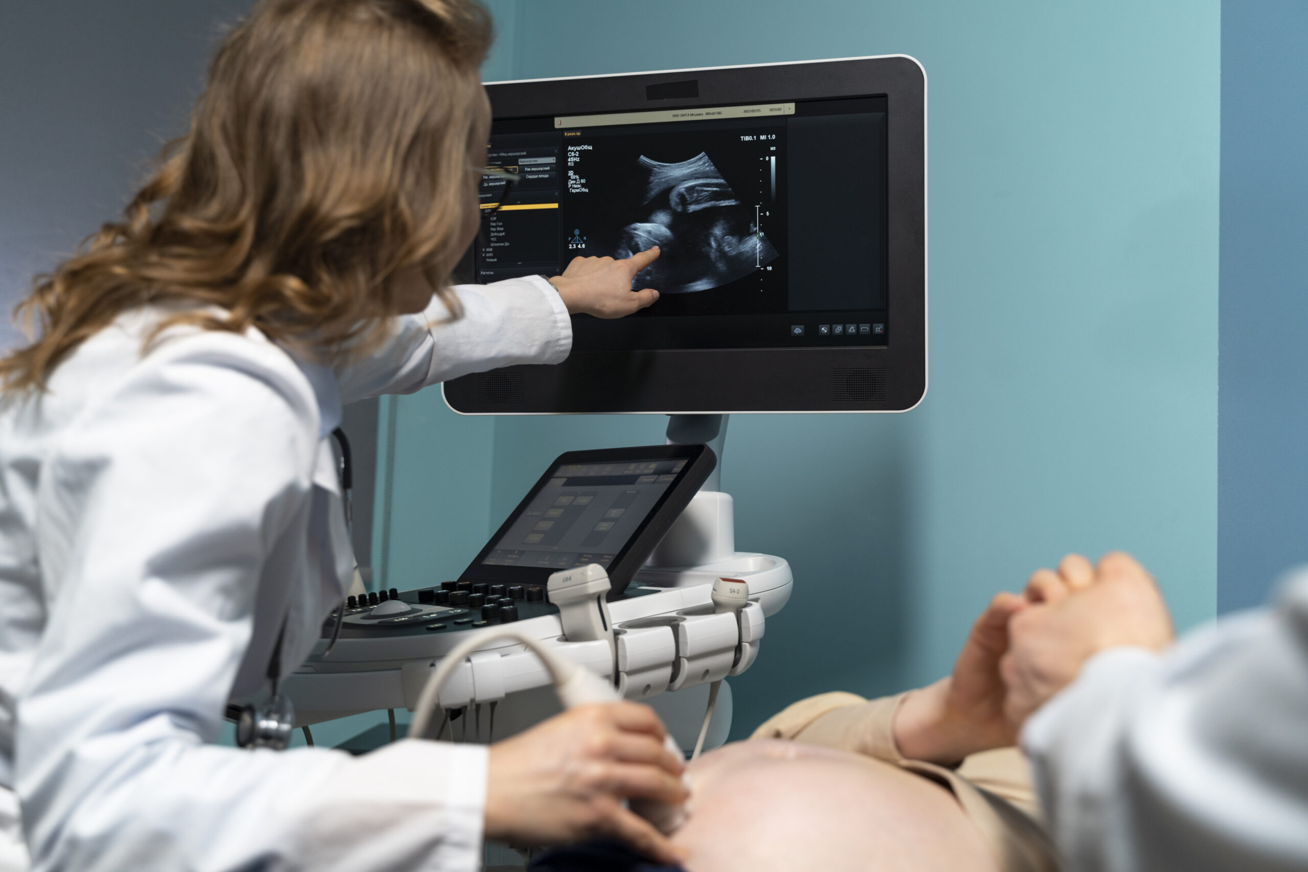
Ultrasound scans are invaluable tools in assessing the health and development of a pregnancy. Two primary types of scans are commonly used:
- Trans-Vaginal Scan (Internal Scan): Typically performed before 11 weeks, this method involves placing a probe in the vagina for a clearer and more accurate early pregnancy picture.
- Trans-Abdominal Scan: From 11 or 12 weeks onwards, a trans-abdominal scan becomes more common. A special gel is applied to the lower abdomen, and a scanner is moved over the area to visualize the uterus and pregnancy.
Routine Ultrasound Scans and Additional Checks:
Routine ultrasound scans between 11 and 14 weeks are essential for:
- Confirming a heartbeat
- Assessing size and growth
- Estimating delivery date
- Identifying the number of babies
Some individuals may be offered a nuchal scan between 11 and 14 weeks to detect chromosomal abnormalities. Hospitals may also provide an anomaly scan at 18-14 weeks for a detailed check of the baby’s development.
Other Possible Scan Findings:
In addition to assessing the viability of the pregnancy, ultrasound scans may reveal other critical information that shapes the course of your pregnancy journey:
Ectopic Pregnancy:
An ectopic pregnancy occurs when the embryo implants outside the uterus, often in one of the Fallopian tubes. Immediate medical attention is necessary due to the risk it poses to maternal health.
Molar Pregnancy:
Placental cells grow rapidly in a molar pregnancy, preventing the baby from developing. Timely medical consultation is essential for appropriate management. Timely medical consultation is essential for appropriate management.
Anatomic Abnormalities and Recurrent Pregnancy Loss (RPL):
Anatomic abnormalities contribute to 10% to 15% of cases of Recurrent Pregnancy Loss (RPL). These abnormalities, such as congenital uterine anomalies, intrauterine adhesions, and uterine fibroids or polyps, can disrupt the vascular supply of the endometrium, leading to abnormal placentation and an increased risk of miscarriage.
Ultrasound plays a pivotal role in identifying anatomic abnormalities associated with Recurrent Pregnancy Loss (RPL). These abnormalities, which consist of congenital uterine anomalies, intrauterine adhesions, and uterine fibroids or polyps, disrupt the vasculature of the endometrium, leading to abnormal and inadequate placentation.
Congenital uterine anomalies, like the uterine septum, are closely associated with Recurrent Pregnancy Loss (RPL). Various Müllerian anomalies, such as unicornuate, didelphic, and bicornuate uteri, also connect to different levels of increased risk in RPL The role of the arcuate uterus in causing RPL remains unclear.
Intrauterine adhesions, often related to Asherman syndrome, can significantly impact placentation, potentially resulting in early pregnancy loss. Larger intramural fibroids and submucosal fibroids are additional factors that may contribute to RPL. Understanding these anatomic abnormalities through ultrasound assessments is crucial for personalized care and informed decision-making in cases of recurrent pregnancy loss. Consultation with healthcare providers allows for a comprehensive evaluation and tailored management strategies based on individual circumstances.